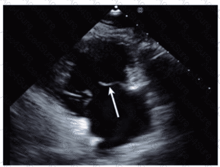
Which anatomic structure is represented by the arrow on this image?
A.
Left leaflet
B.
Posterior leaflet
C.
Septal leaflet
D.
D. Anterior leaflet
The Answer Is:
D
This question includes an explanation.
Explanation:
The echocardiographic image is a four-chamber view focusing on the mitral valve apparatus. The arrow points to the anterior leaflet of the mitral valve, which is typically more prominent, triangular in shape, and located adjacent to the aortic valve in the left ventricular outflow tract region.
The posterior leaflet of the mitral valve is generally smaller, has multiple scallops, and is located posteriorly relative to the anterior leaflet. The septal leaflet is part of the tricuspid valve on the right side of the heart. The "left leaflet" is a non-specific term and not an anatomical descriptor.
This differentiation between anterior and posterior leaflets is important for understanding mitral valve pathology and for interventions such as mitral valve repair. These features are clearly explained in echocardiography texts and ASE valve imaging guidelines【12:ASE Valve Imaging Guidelines†p.180-185】【16:Textbook of Clinical Echocardiography, 6e†p.200-205】.
AE-Adult-Echocardiography PDF/Engine
Printable Format
Value of Money
100% Pass Assurance
Verified Answers
Researched by Industry Experts
Based on Real Exams Scenarios
100% Real Questions
Get 60% Discount on All Products,
Use Coupon: "8w52ceb345"