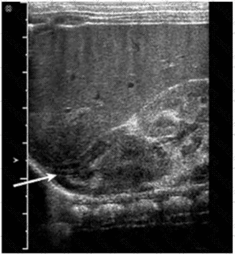
The structure indicated by the arrow in the ultrasound image is the adrenal gland. On ultrasound, the adrenal gland in neonates and infants is relatively large and has a distinctive “Y” or “V” shape in the transverse view. It is located superior and slightly medial to the upper pole of the kidney.
In this image, the arrow is pointing to a hypoechoic, curved structure with a thin echogenic central stripe, representing the fetal adrenal gland. This echogenic stripe corresponds to the adrenal medulla, while the surrounding hypoechoic area represents the cortex.
Differential features:
A. Kidney: While the kidney is visualized posterior to the adrenal gland and shows a reniform shape with a central echogenic sinus and peripheral cortex, it is not the structure being directly pointed to by the arrow.
B. Bowel loop: Bowel has variable echogenicity with peristalsis and shadowing from air. It does not have the consistent morphology or location seen in the image.
C. Diaphragm: Appears as a thin, hyperechoic linear structure separating the thoracic cavity from the abdomen. It is seen more superiorly than the indicated structure and lacks the “Y” or “V” adrenal configuration.
Key Anatomical Landmarks:
The adrenal glands are located in the retroperitoneum, superior to the kidneys, and appear prominent on ultrasound in neonates.
In transverse view, the right adrenal gland is anterior to the crus of the diaphragm and posterior to the inferior vena cava (IVC).
[References:, Rumack CM, Wilson SR, Charboneau JW, Levine D. Diagnostic Ultrasound. 5th Edition. Elsevier, 2018. Chapter: Adrenal Glands and Retroperitoneum, pp. 291–295., American Institute of Ultrasound in Medicine (AIUM) Practice Parameter for the Performance of an Ultrasound Examination of the Abdomen and/or Retroperitoneum. 2020., , , ]