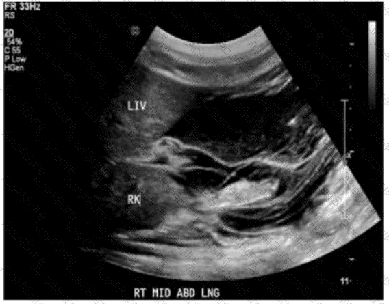
The ultrasound image shows an anechoic (black) fluid collection in the perihepatic and perirenal spaces. The fluid outlines the liver (LIV) and right kidney (RK), which is characteristic of free fluid in the peritoneal cavity — consistent with ascites.
Sonographic features of ascites:
Anechoic (or hypoechoic) fluid in dependent areas of the abdomen
Seen surrounding the liver, spleen, and intestines
Can be free-flowing or loculated
Bowel loops may be floating or displaced centrally
This image is consistent with a typical finding of ascites: free fluid in Morison’s pouch (hepatorenal recess), a common site for fluid accumulation.
Differentiation from other options:
A. Hydropic gallbladder: Refers to an enlarged gallbladder filled with clear bile; not visible in this image.
B. Hemoperitoneum: May appear similar to ascites, but usually has complex echogenicity or layering if acute; clinical context (trauma, bleeding) is essential for diagnosis.
C. Bowel obstruction: Would show dilated, fluid-filled bowel loops with peristalsis or to-and-fro motion, not evident here.
[References:, Rumack CM, Wilson SR, Charboneau JW, Levine D. Diagnostic Ultrasound. 5th Edition. Elsevier, 2018. Chapter: Peritoneal Cavity and Abdominal Trauma, pp. 125–130., American Institute of Ultrasound in Medicine (AIUM). Practice Parameter for the Performance of a Focused Assessment with Sonography for Trauma (FAST) Examination, 2020., Radiopaedia.org. Ascites (ultrasound): https://radiopaedia.org/articles/ascites-ultrasound, ]